Surgical Technique for OrthoACITM
OrthoACITM Product Description
- OrthoACITM is an autologous cell therapy manufactured by in vitro expansion of chondrocytes harvested from the patient’s own healthy articular cartilage. Expanded chondrocytes are loaded onto a collagen scaffold and implanted into the cartilage defect.
- Orthocell’s OrthoACITM is indicated for use in the treatment of cartilage defects associated with the knee, patella and ankle.1.
- Patient selection criteria include:
- Patients with symptomatic cartilage damage caused by trauma, wear or degradation including International Cartilage Repair Society (ICRS) grade III or IV cartilage lesions associated with chondromalacia patella or osteochondritis dissecans.
- Patients in the age range of 18 – 55 years.
- OrthoACITM is approved by the Therapeutic Goods Administration (TGA) and is listed on the Australian Register of Therapeutic Goods (ARTG).2.
01
OrthoACITM First Stage – Biopsy3.
Biopsy Procedure
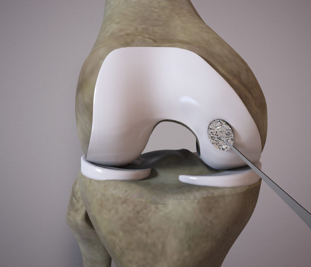
- Take the biopsy from the articular cartilage in a non-weight bearing region (Fig. 1A).
- The size of the biopsy should ideally be 3-6mm long or 200-300mg of cartilage.
- Place the biopsy in the transport medium supplied and replace the lid securely.
- Collect blood samples at the time of biopsy (75ml) and place into the tubes provided.
- The cell expansion process takes between 4 and 6 weeks before the cells are ready to be implanted.
 1A
1A - For the full list of indications and contraindications, please refer to OrthoACITM Product Information.
- TGA Licence no. MI-19052008-002420-11; ARTG number 289402.
- Please refer to OrthoACITM Receipt of the Biopsy Pack and OrthoACITM Use of the Biopsy Pack.
02
OrthoACITM Second Stage – Site Preparation
Biopsy Procedure
- Healthy cartilage margins are required to facilitate the integration and regeneration process following the implant.
- Circumscribe the defect with a scalpel or curette to expose healthy cartilage, leaving a vertical cartilage wall/face.
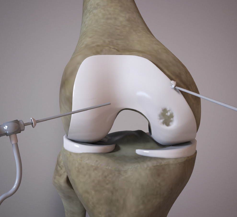
- Debride the damaged cartilage down to, but not through, the subchondral bone plate using a ring curette or similar instrument (Fig. 2A)
 2A
2A 03
OrthoACITM Second Stage – Cell Loading
 3A
3A 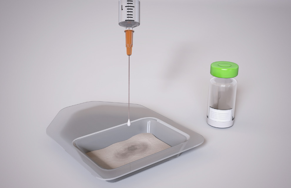 3B
3B Prior to the procedure starting, the theatre support staff load the autologous chondrocytes on to the rough side of the supplied collagen scaffold.
Drawing Cells
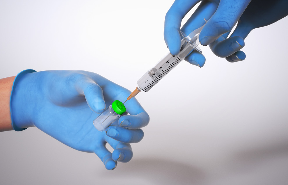
- Cells must be resuspended prior to use.
- Invert the vial gently several times until a uniform suspension is formed.
- Remove the centre aluminium tab from the green crimp seal and wipe with an alcohol swab
- Use a 19 gauge needle with a 3ml syringe to gently draw up the cell suspension (Fig. 3A).
Biopsy Procedure
- Gently load the cells on to the rough side of the collagen scaffold, ensuring that the entire surface area of the scaffold is evenly covered with the fluid containing the cells (Fig. 3B).
- 2 vials of OrthoACITM is adequate for a 30x40mm scaffold.
- The cells are left on the scaffold for a minimum of 20 minutes prior to implant.
04
OrthoACITM Second Stage – Collagen Scaffold Sizing
 4A
4A 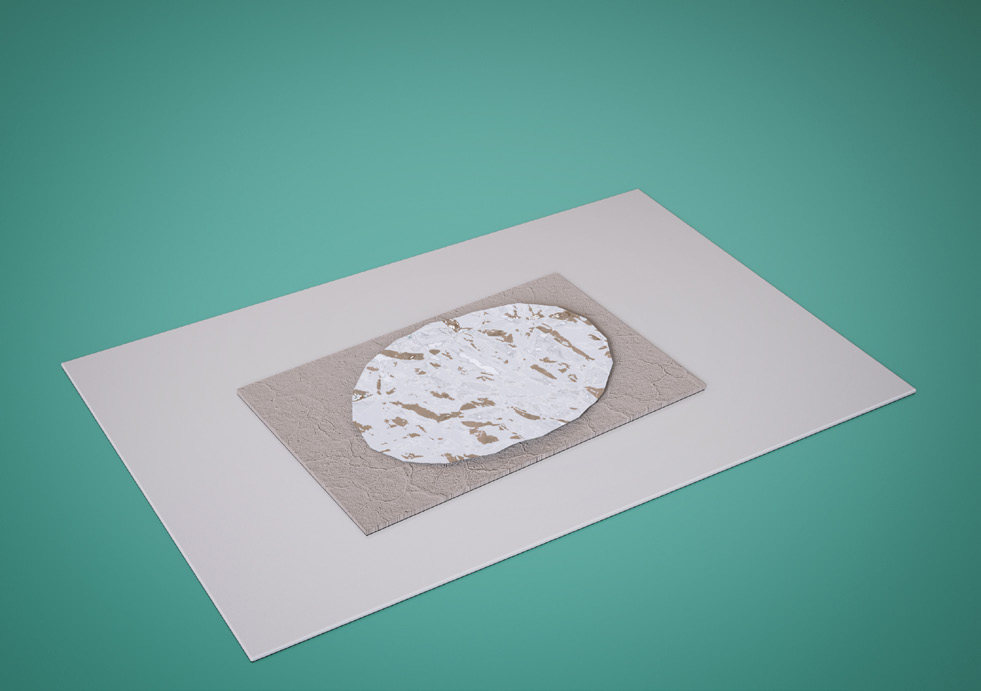 4B
4B 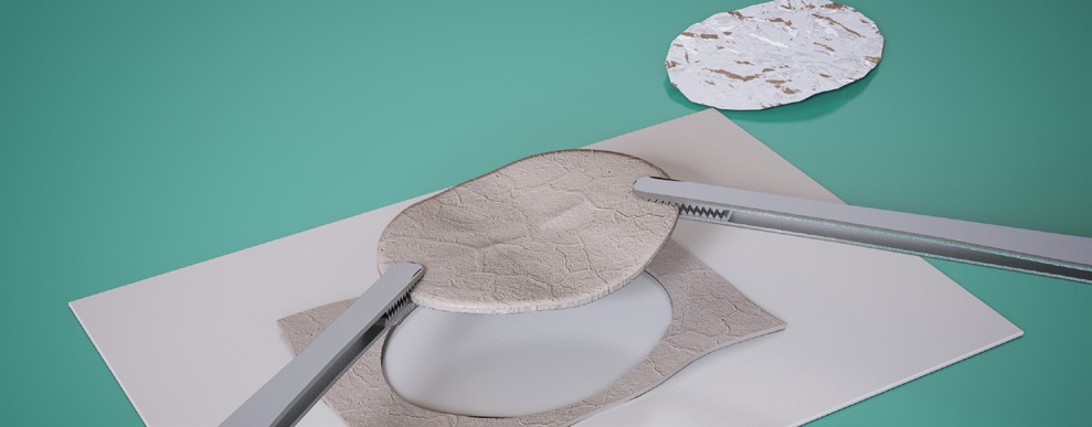 4C
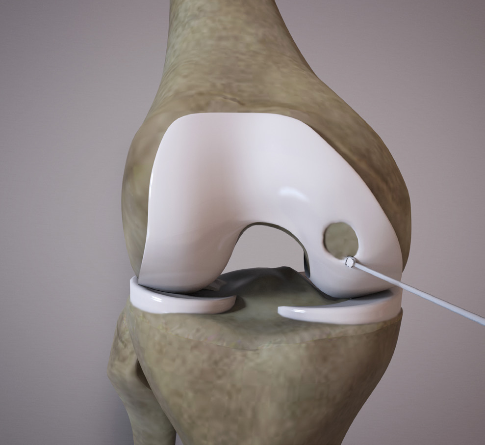
4C - Use a sterile foil template to make an impression of the defect (Fig. 4A).
- Place the foil template on the rough side of the scaffold and cut to size with scissors (Fig. 4B and 4C).
05
OrthoACITM Second Stage – Collagen Scaffold Implant
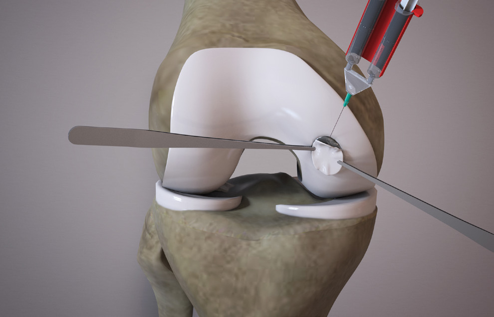 5A
5A 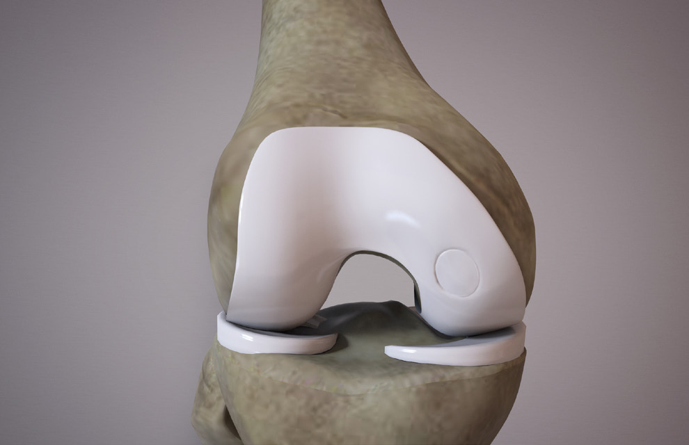 5B
5B - Place the OrthoACITM loaded scaffold rough side down, facing the subchondral bone plate.
- Retract the scaffold and apply a fibrin sealant to the subchondral bone plate (Fig. 5A).
- Seal the OrthoACITM loaded scaffold into position by applying gentle pressure for 1 minute.
- Remove excess glue and overhanging scaffold.
- Put the joint through a range of motion to ensure that the graft is securely in position (Fig. 5B).
Post Operation Rehabilitation4.
- A prescribed rehabilitation program is recommended to support optimal clinical outcomes. 5.
- OrthoACITM Rehabilitation Guide: Ankle or OrthoACITM Rehabilitation Guide: Knee/Patella.
- A Randomized Trial Comparing Accelerated and Traditional Approaches to Postoperative Weightbearing Rehabilitation After Matrix-Induced Autologous. Chondrocyte Implantation: Findings at 5 Years – Am J Sports Med. 2012 Jul;40(7):1527-37. Ebert JR, Fallon M, Zheng MH, Wood DJ, Ackland TR.
Preparation for OrthoACITM Biopsy 6.
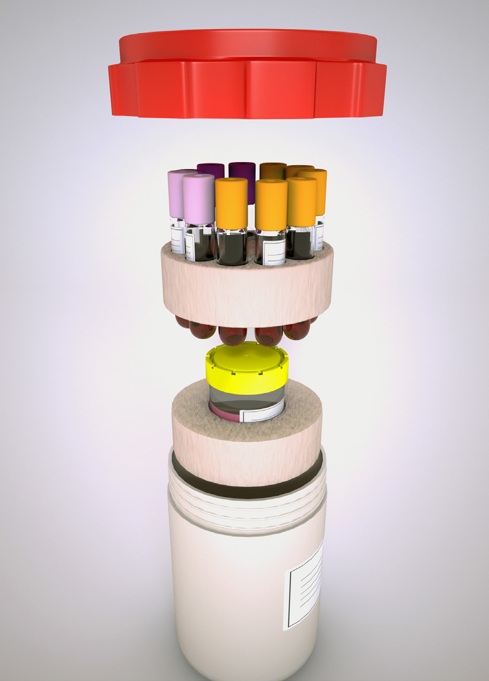
- On receiving the OrthoACITM Biopsy Kit, place transport container [Fig. 6A] in fridge and ice packs in freezer until time of biopsy.
- IMPORTANT – all documentation must be completed and signed by the patient and doctor.
Biopsy Sample Collection and Packing of Biopsy Kit for Transport 7.
- Collect blood samples at the time of biopsy (75ml) in the tubes provided.
- Place the biopsy and blood tubes in foam supports.
- Place all samples in the plastic bag provided and seal with the tamper-proof label (red) provided.
- Place bagged samples into the Transport Container and close [Fig. 6A].
- IMPORTANT – all specimens (biopsy in transport medium and blood tubes) must be correctly labelled with patient information. Unlabelled specimens will be discarded.
 2A
2A Implant Receipt Instructions 8.
- DO NOT refrigerate the Implant Kit.
- The Implant Kit must be kept sealed at room temperature until time of procedure.
- Product expiry is 72 hours from time of preparation.
- Expiry information is indicated on all product labels, including the product certificate.
- The transport container holds a minimum of two vials, each containing 1ml of chondrocyte cell suspension.
- According to the defect size(s), Orthocell will supply the required amount of vials and scaffold for a truly customised ACI procedure.
Key Clinical Data for ACI Therapies
- Matrix-Applied Characterized Autologous Cultured Chondrocytes Versus Microfracture: Two-Year Followup of a Prospective Randomized Trial Am J Sports Med. 2014 42(6):1384-94. Saris D et al.
- Matrix-Applied Characterized Autologous Cultured Chondrocytes Versus Microfracture Five-Year Followup of a Prospective Randomized Trial Am J Sports Med. 2018 46(6):1343-51. Brittberg M, Recker D, Ilgenfritz J & Saris DBF
- Matrix-Associated Autologous Chondrocyte Implantation: A Clinical Follow-Up at 15 Years Cartilage 2016 Oct;7(4):309-15. Gille J, Behrens P, Schulz AP, Oheim R & Kienast B
- Minimum 10-Year Clinical and Radiological Outcomes of a Randomized Controlled Trial Evaluating 2 Different Approaches to Full Weight-bearing After Matrix-Induced Autologous Chondrocyte Implantation Am J Sports Med. 2020 48(1):133-142. Ebert JR, Fallon M, Ackland TR, Janes GC & Wood DJ
- Please refer to OrthoACITM Receipt of the Biopsy Pack
- Please refer to OrthoACITM Use of the Biopsy Pack
- Please refer to Receipt and Use of the OrthoACITM Implant Pack